


Eye Anatomy
Eye Anatomy
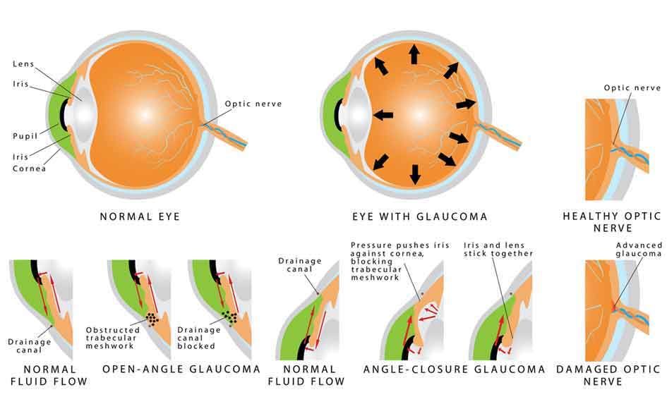
CORNEA: The clear, protective front layer covering the iris, pupil and anterior eye chamber.
PUPIL: The circular black hole located in the center of the iris that regulates the amount of light allowed to enter the eye and strike the retina.
IRIS: The pigmented portion of the eye that is located behind the cornea. It provides eye color and controls incoming light.
LENS: A layer of transparent intraocular tissue that acts as a natural lens to help direct rays of light to the retina.
RETINA: Consisting of many rods and cones that convert the images we see into electrical impulses that get sent through the optic nerve to the brain.
MACULA: A small, centralized section of the retina that manages sharp central vision.
VITREOUS: Colorless, gelatinous filling in the rear interior eyeball, between the lens and the retina.
OPTIC NERVE: The largest sensory nerve that carries impulses for a vision from the retina to the brain.
SCLERA: The visible white portion of the eye. A protective fibrous outer layer that covers most of the eyeball except for the cornea.
CILIARY BODY: A muscular ring beneath the front of the eyeball that aids in focus by changing the lens shape. It also produces aqueous humor.
CHOROID: A layer of blood vessels between the sclera and the retina that help channel oxygen and nutrients to the eye.


Interested in our
Services?
Request an appointment with this form to schedule a time with our professional staff!
Phone: (734) 562-0099
Fax: 734-854-5868
- Monday8:30 am - 7:00 pm
- Tuesday8:30 am - 5:00 pm
- Wednesday11:00 am - 7:00 pm
- Thursday8:30 am - 5:00 pm
- Friday7:30 am - 4:00 pm
- SaturdayClosed
- SundayClosed
Powered by: 